
Manual Resuscitation: A Comprehensive Overview (Updated February 15, 2026)
This detailed guide explores essential manual resuscitation techniques, incorporating the latest 2023 AHA/AAP neonatal updates focusing on improved newborn care and outcomes.
Manual resuscitation encompasses the life-saving interventions performed before the availability of advanced medical technology. It’s a cornerstone of emergency care, relying on fundamental skills like airway management, breathing support, and chest compressions to maintain circulation and oxygenation. This approach is crucial in situations where immediate intervention can dramatically improve patient outcomes, particularly in pre-hospital settings or during the initial moments of a cardiac or respiratory arrest.
Effective manual resuscitation requires consistent training and adherence to established guidelines, such as those provided by the American Heart Association (AHA) and the American Academy of Pediatrics (AAP). Recent updates, like the 2023 focused update to neonatal resuscitation guidelines, emphasize optimizing techniques for vulnerable populations, like newborns, focusing on areas like umbilical cord management and appropriate ventilatory support. Mastering these skills empowers individuals to become vital links in the chain of survival.
The Importance of Early Intervention
The critical nature of early intervention in resuscitation cannot be overstated. Every minute without oxygenated blood flow drastically reduces the chances of survival and increases the risk of irreversible brain damage. Initiating manual resuscitation techniques – even before professional help arrives – buys valuable time, maintaining vital organ function and maximizing the potential for a positive outcome.
Prompt action is particularly crucial in neonatal cases, as highlighted by the recent AHA/AAP updates. Effective umbilical cord management and immediate ventilatory support can significantly impact a newborn’s transition to extrauterine life. Delays in these initial steps can lead to severe complications. Early, consistent, and correct application of manual resuscitation skills represents the best opportunity to prevent morbidity and mortality, emphasizing the power of immediate response.

Assessing the Patient
Rapid and accurate patient assessment is paramount, beginning with the ABCs – Airway, Breathing, and Circulation – to guide effective resuscitation efforts.
Initial Assessment: ABCs (Airway, Breathing, Circulation)
The initial assessment prioritizes the ABCs – Airway, Breathing, and Circulation – forming the cornerstone of effective manual resuscitation. First, ensure a patent Airway; look for obstructions and prepare to clear them. Next, assess Breathing: observe chest rise, listen for breath sounds, and determine respiratory rate and effort. Is the patient breathing adequately?
Simultaneously, evaluate Circulation by checking for a pulse – carotid in adults and infants, brachial in infants – and assessing skin color, temperature, and capillary refill time. These vital signs provide immediate insight into the patient’s condition. A compromised airway, absent breathing, or lack of pulse necessitates immediate intervention. This systematic approach allows rescuers to quickly identify life-threatening issues and prioritize interventions, maximizing the chances of a successful resuscitation.
Checking for Responsiveness
Determining responsiveness is the crucial first step in any resuscitation effort. Begin by gently stimulating the patient, using both verbal and tactile cues. Vigorously shout, “Are you okay?” while simultaneously tapping or shaking the shoulders. Observe for any signs of response – movement, eye-opening, or verbalization.
If there’s no response, proceed immediately to activate the emergency response system (call for help). A lack of responsiveness indicates a potential loss of consciousness and requires prompt intervention. It’s vital to avoid unnecessary movement of the patient’s head and neck, especially if trauma is suspected. Document the absence of response and the time it was noted, as this information is critical for subsequent care and accurate record-keeping during the resuscitation process.
Pulse Assessment & Location
Accurate pulse assessment is fundamental to evaluating circulation during resuscitation. In adults, the carotid pulse – located in the neck, beside the trachea – is the primary site for quick assessment; In infants and children, the brachial pulse, found on the inner aspect of the upper arm, is preferred due to its easier palpation.
Palpate gently but firmly, using the pads of your fingers, avoiding the trachea itself. Note the rate, rhythm, and quality of the pulse. A weak or absent pulse indicates inadequate perfusion and necessitates immediate initiation of chest compressions. Remember that pulse assessment should be brief – no more than 10 seconds – to minimize interruptions to compressions if they are needed. Document findings clearly.

Airway Management
Establishing and maintaining a patent airway is paramount; techniques like head-tilt/chin-lift and jaw-thrust maneuvers are crucial for effective oxygenation.
Opening the Airway: Head-Tilt/Chin-Lift & Jaw-Thrust
Opening the airway is the first critical step in resuscitation. The head-tilt/chin-lift maneuver is commonly used when there’s no suspicion of cervical spine injury. Gently tilting the head back and lifting the chin helps lift the tongue away from the posterior pharynx, creating an open airway.
However, if a cervical spine injury is suspected, the jaw-thrust maneuver is preferred. This technique involves grasping the angles of the mandible and lifting it forward, avoiding any head extension. This minimizes movement of the cervical spine.
Proper technique is vital; excessive force can cause injury. Assess for any obstructions after attempting either maneuver. Both methods aim to relieve the tongue’s obstruction and facilitate airflow, ensuring effective ventilation during resuscitation efforts. Regular reassessment is key.
Suctioning Techniques for Airway Clearance
Effective airway clearance is paramount during resuscitation, and suctioning plays a crucial role in removing obstructions like secretions, blood, or foreign bodies. Utilize a rigid tonsil tip suction catheter (Yankauer) for visible obstructions within the oropharynx. Insert the catheter gently, applying suction only during withdrawal to avoid trauma.
For infants, a flexible suction catheter is often preferred. Limit suctioning to 10 seconds per pass to prevent hypoxia. Always pre-oxygenate the patient before and after suctioning, if possible.
Proper suctioning technique minimizes airway trauma and maximizes oxygenation. Ensure the suction device is functioning correctly and maintain sterile technique. Frequent bulb syringe suctioning is vital for newborns.
Foreign Body Airway Obstruction (FBAO) – Adult
Recognizing and swiftly addressing adult FBAO is critical. If a conscious adult exhibits signs of choking – inability to speak, cough, or breathe – initiate the Heimlich maneuver immediately. Stand behind the victim, wrap your arms around their waist, and make a fist with one hand.
Place the thumb side of your fist against the victim’s abdomen, slightly above the navel and below the ribcage. Grasp your fist with your other hand and deliver quick, upward thrusts. Continue until the object is dislodged or the victim becomes unresponsive.
If the victim loses consciousness, carefully lower them to the ground and begin CPR, checking for the object before each ventilation attempt.
Foreign Body Airway Obstruction (FBAO) – Infant
Infant FBAO requires a modified approach. Support the infant face down along your forearm, ensuring head support. Deliver five firm back blows between the shoulder blades using the heel of your hand. If unsuccessful, carefully turn the infant face up, supporting the head and neck.
Place two fingers in the center of the infant’s chest, just below the nipple line, and deliver five quick chest thrusts, compressing approximately 1.5 inches. Alternate between five back blows and five chest thrusts until the object is dislodged or the infant becomes unresponsive.
If unresponsiveness occurs, begin infant CPR, checking the mouth for the obstructing object before each ventilation.

Breathing Support
Effective breathing support involves rescue breathing and Bag-Valve-Mask (BVM) ventilation, crucial for oxygenating patients unable to breathe adequately on their own.
Rescue Breathing Techniques
Rescue breathing provides vital oxygen when a patient isn’t breathing, or isn’t breathing effectively. Proper technique is paramount for successful ventilation. Begin by ensuring the airway is open, utilizing head-tilt/chin-lift or jaw-thrust maneuvers as appropriate. Pinch the patient’s nostrils closed, creating a tight seal over their mouth with yours.
Deliver breaths over one second each, observing for visible chest rise. Avoid excessive force or volume, as this can cause gastric inflation. Allow for full chest recoil between breaths. The rate of rescue breathing depends on the situation; for adults, typically 10-12 breaths per minute, and for infants and children, a slightly faster rate is often needed. Continuous assessment of breathing effectiveness is crucial, adjusting technique as necessary to maintain adequate oxygenation until spontaneous breathing resumes or advanced airway management is implemented.
Bag-Valve-Mask (BVM) Ventilation
Bag-Valve-Mask (BVM) ventilation is a critical skill for providing assisted ventilation when spontaneous breathing is inadequate. Select an appropriately sized mask ensuring a tight seal over the patient’s mouth and nose. Utilize the ECIN technique – Ensure Chest Rise, Compress the bag, Inhale over one second, and Note chest movement.
Maintain a firm two-handed grip, squeezing the bag to deliver breaths while observing for visible chest rise. Avoid excessive ventilation, which can lead to complications. Proper technique requires training and practice. BVM ventilation is often used in conjunction with airway adjuncts to optimize airway patency. Continuous monitoring of oxygen saturation and ventilation effectiveness is essential, adjusting the rate and volume as needed to maintain adequate oxygenation and ventilation.
Ventilation Rate and Volume
Determining appropriate ventilation rate and volume is crucial for effective manual resuscitation. For adults, a ventilation rate of 10-12 breaths per minute is generally recommended, delivering each breath over one second. Aim for visible chest rise with each breath, indicating adequate tidal volume – typically around 6-7 ml/kg of ideal body weight.
In infants and children, ventilation rates are higher, ranging from 20-30 breaths per minute. Careful observation of chest movement is paramount; avoid over-inflation, which can cause lung injury. Adjust ventilation based on patient response, monitoring oxygen saturation and end-tidal CO2 levels when available. Consistent, controlled breaths are more important than achieving a specific volume.
Circulation & Chest Compressions
Effective chest compressions and circulation restoration are vital components of manual resuscitation, ensuring oxygenated blood reaches vital organs promptly and efficiently.
Chest Compression Technique – Adult
Adult chest compressions require proper hand placement and technique for optimal effectiveness. Position the heel of one hand in the center of the patient’s chest, on the lower half of the sternum. Place the other hand on top of the first, interlacing fingers to avoid applying pressure to the ribs.
Maintain a straight-arm position, locking elbows and using body weight to deliver compressions. Depress the chest at least 2 inches (5 cm), but no more than 2.4 inches (6 cm). Allow for complete chest recoil after each compression, enabling the heart to refill with blood.
Minimize interruptions to compressions, as continuous flow is crucial. Effective compressions generate palpable arterial pulses and visible chest rise, indicating adequate circulation. Remember, high-quality compressions are the cornerstone of successful resuscitation efforts.
Chest Compression Technique – Infant
Infant chest compressions differ from adult technique due to the smaller chest size and bone structure. Utilize two fingers (index and middle) or two thumbs encircling the chest, positioned just below the nipple line on the lower half of the sternum.
Compress the chest approximately 1.5 inches (4 cm), ensuring complete chest recoil after each compression. Maintain a compression rate of 100-120 compressions per minute. Avoid excessive force, as infants are more susceptible to injury.
Proper hand placement and controlled depth are vital for effective circulation. Continuous compressions, minimizing interruptions, are paramount. Assess for signs of circulation, such as palpable pulses or visible chest rise, while delivering compressions.
Compression Depth and Rate
Maintaining appropriate compression depth and rate is crucial for effective manual resuscitation. For adults, compress the chest at least 2 inches (5 cm), but no more than 2.4 inches (6 cm). Infants require a compression depth of approximately 1.5 inches (4 cm).
The recommended compression rate for both adults and infants is 100-120 compressions per minute. This rapid pace helps to circulate blood and oxygen to vital organs. Allow for complete chest recoil between each compression to facilitate venous return.
Consistent depth and rate are essential; avoid leaning on the chest or compressing too slowly. Utilize a metronome or memory aid to maintain the correct rhythm throughout the resuscitation effort.
The 30:2 Compression-to-Ventilation Ratio
The established 30:2 compression-to-ventilation ratio is a cornerstone of effective manual resuscitation for adults. This means delivering 30 chest compressions followed by two rescue breaths. This cycle maximizes oxygen delivery while minimizing interruptions to chest compressions, which are vital for maintaining circulation.
This ratio applies when a single rescuer is performing CPR. With two or more rescuers, a compression-to-ventilation ratio of 30:2 is still recommended, but allows for continuous compressions with ventilation provided every 6 seconds.
Adhering to this ratio ensures a balance between mechanical circulation and oxygenation, improving the chances of successful resuscitation. Consistent application is key to optimal patient outcomes.

Neonatal Resuscitation Specifics
Newborn resuscitation demands specialized techniques, including careful umbilical cord management and tailored ventilatory support, as per the latest AHA/AAP guidelines.
Umbilical Cord Management
Optimal umbilical cord management is crucial during neonatal resuscitation, significantly impacting newborn transition and outcomes. Current guidelines, updated by the AHA and AAP in 2023, emphasize a shift away from immediate cord clamping in many cases. Delayed cord clamping – waiting 30 to 60 seconds after birth – allows for continued placental transfusion, providing the infant with vital blood volume and iron stores.
However, this approach requires careful consideration. If the newborn exhibits signs of respiratory distress or requires immediate resuscitation, clamping and cutting the cord should not be delayed. The focus remains on prioritizing the infant’s respiratory and circulatory needs. Healthcare providers must be proficient in assessing the newborn’s condition and making informed decisions regarding cord management, balancing the benefits of delayed clamping with the necessity for rapid intervention when indicated.
Ventilatory Support in Newborns
Effective ventilatory support is paramount in neonatal resuscitation when a newborn struggles to establish spontaneous breathing. The 2023 AHA/AAP guidelines highlight the importance of initiating positive-pressure ventilation (PPV) promptly when a newborn exhibits signs of respiratory distress, such as gasping or apnea. PPV delivers air or oxygen to the lungs, assisting with chest expansion and oxygenation.
Techniques include bag-valve-mask (BVM) ventilation, requiring proper mask seal and appropriate ventilation rate. Initial breaths should be delivered within the first few seconds of life, aiming for a heart rate increase. Careful monitoring of chest rise and auscultation are essential to avoid over-ventilation, which can cause lung injury. The guidelines emphasize individualized approaches based on the newborn’s gestational age and clinical condition.
Neonatal Resuscitation Guidelines (AHA/AAP 2023 Update)
The American Heart Association (AHA) and American Academy of Pediatrics (AAP) released a focused update to their 2020 neonatal resuscitation guidelines in 2023. This update centers on optimizing initial steps and refining ventilatory support strategies for newborns requiring assistance at birth. Key changes emphasize earlier and more effective positive-pressure ventilation (PPV) when spontaneous breathing is inadequate.
Significant attention is given to umbilical cord management, advocating for delayed clamping in stable newborns to enhance blood volume and iron stores. The guidelines also reinforce the importance of continuous assessment and adaptation of resuscitation efforts based on the infant’s response. These updates aim to improve outcomes and standardize care for newborns needing resuscitation, reflecting current evidence-based practices.

Advanced Considerations
Continuous monitoring of the patient’s response is crucial, determining the need for escalation to advanced life support interventions and specialized medical expertise.
Monitoring Response to Resuscitation
Effective monitoring is paramount throughout manual resuscitation efforts. Regularly assess for signs of returning spontaneous circulation (ROSC), including purposeful movement, coughing, or normal breathing patterns. Continuous observation of the patient’s clinical status provides vital feedback on the efficacy of interventions.
Key indicators to monitor include changes in skin perfusion – noting color and temperature – and improvements in respiratory effort. Pulse oximetry, when available, can help gauge oxygen saturation levels. However, reliance solely on numerical data is insufficient; clinical judgment remains essential.
Frequent reassessments of the ABCs (Airway, Breathing, Circulation) are necessary to identify any deterioration or lack of improvement. Document all observations and interventions meticulously, facilitating clear communication within the resuscitation team and informing subsequent decisions regarding continued manual resuscitation or transition to advanced life support.
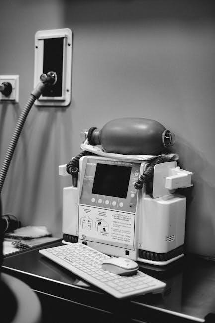
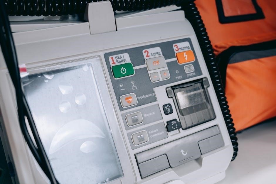
When to Transition to Advanced Life Support
Despite diligent manual resuscitation, certain scenarios necessitate a swift transition to advanced life support (ALS). Prolonged absence of ROSC – return of spontaneous circulation – after a reasonable period of high-quality CPR is a primary indicator. This timeframe should be determined by established protocols and experienced clinical judgment.

Persistent ventricular fibrillation or pulseless ventricular tachycardia, unresponsive to initial defibrillation attempts, also warrants escalation to ALS. Similarly, if the patient’s condition deteriorates despite ongoing manual interventions, or if underlying reversible causes are suspected but cannot be addressed with basic measures, ALS becomes crucial.
Early involvement of ALS resources maximizes the chances of successful resuscitation. A seamless handover, providing a concise report of interventions and patient response, is vital for optimal care continuation.