Fluoro-Guided Injections: A Comprehensive Overview (Updated February 15, 2026)
Fluoroscopy-free RIRS demonstrates success, yet fluoroscopy remains crucial for managing unforeseen difficulties during surgery, ensuring optimal patient outcomes and safety.
What are Fluoro-Guided Injections?
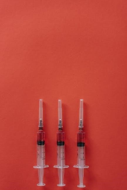
Fluoro-guided injections are minimally invasive procedures utilizing fluoroscopy – a type of real-time X-ray – to precisely guide the placement of a needle into a specific anatomical location. This technique allows physicians to visualize the needle’s trajectory and confirm accurate delivery of medication, such as corticosteroids or local anesthetics, directly to the targeted area.
Unlike traditional “blind” injections, where the physician relies on anatomical landmarks, fluoroscopy provides a dynamic visual confirmation. This is particularly valuable for deep-seated structures or areas where anatomical variations are common. The use of fluoroscopy enhances accuracy, maximizing therapeutic benefit and minimizing the risk of unintended consequences. It’s a cornerstone of modern interventional pain management and diagnostic procedures.
The Role of Fluoroscopy in Injection Procedures
Fluoroscopy plays a pivotal role by providing real-time visualization during injection procedures, acting as the “eyes” of the physician. It allows for precise needle guidance, ensuring accurate placement within the intended anatomical structure – be it a joint, nerve root, or other target. This dynamic imaging confirms the needle reaches the desired location before medication is delivered.
Furthermore, fluoroscopy aids in verifying appropriate medication spread, confirming it reaches the intended area and avoids unintended diffusion. It’s especially crucial when anatomical landmarks are obscured or variations exist. While fluoroscopy-free techniques are emerging, its availability remains vital for addressing unexpected intraoperative challenges, ensuring optimal patient safety and treatment efficacy.
Why Use Fluoroscopic Guidance?

Fluoroscopic guidance dramatically enhances the accuracy and safety of injections, minimizing the risk of complications. It’s particularly valuable when targeting deep-seated structures or those with complex anatomy where palpation alone is insufficient. Real-time visualization confirms precise needle placement, reducing the chance of injecting into unintended tissues or missing the target altogether.
Moreover, fluoroscopy allows for confirmation of appropriate medication distribution, ensuring the therapeutic agent reaches the intended site for maximum benefit. Even with emerging fluoroscopy-free techniques, the ability to quickly adapt and address unexpected challenges during a procedure remains paramount, making fluoroscopy a crucial safety net for both physician and patient.
Common Applications of Fluoro-Guided Injections
Fluoro-guided injections find widespread application across various medical specialties, primarily in musculoskeletal and pain management. Spinal injections, including facet joint, epidural steroid, and sacroiliac joint injections, frequently benefit from fluoroscopic precision to ensure accurate needle placement within the often-complex spinal structures.
Peripheral joint injections, such as those targeting the knee, shoulder, and hip, also utilize fluoroscopy to confirm intra-articular placement, maximizing therapeutic effect. While advancements explore fluoroscopy-free options like in RIRS, the technique remains vital for complex cases. Ultimately, fluoroscopic guidance expands the range of treatable conditions and improves patient outcomes across a broad spectrum of applications.
Spinal Injections
Spinal injections represent a significant portion of fluoro-guided procedures, addressing a wide range of pain conditions affecting the back and neck. Fluoroscopy’s real-time imaging capability is paramount in navigating the intricate anatomy of the spine, ensuring accurate needle positioning for optimal therapeutic delivery.
Specifically, procedures like facet joint injections, epidural steroid injections, and sacroiliac joint injections rely heavily on fluoroscopic guidance. This precision minimizes the risk of complications and maximizes the effectiveness of the injected medication. While research explores alternatives, fluoroscopy remains a cornerstone for achieving successful outcomes in spinal pain management, particularly when unexpected challenges arise during the procedure.
Facet Joint Injections
Facet joint injections, guided by fluoroscopy, are utilized to diagnose and treat pain originating from the small joints in the spine. Fluoroscopic guidance allows physicians to precisely target these joints, confirming accurate needle placement before injecting a local anesthetic and corticosteroid mixture.
This precision is crucial, as misplacement can lead to ineffective pain relief or unintended complications. The real-time visualization provided by fluoroscopy ensures the medication reaches the inflamed facet joint, reducing pain and inflammation. While alternative imaging modalities exist, fluoroscopy’s ability to handle intraoperative challenges makes it a reliable choice for facet joint injections, enhancing diagnostic accuracy and therapeutic efficacy.
Epidural Steroid Injections
Fluoroscopically guided epidural steroid injections are a common treatment for spinal stenosis, herniated discs, and other conditions causing radicular pain. Fluoroscopy ensures accurate needle placement into the epidural space, minimizing the risk of dural puncture or intravascular injection.
The real-time imaging allows physicians to visualize the spread of contrast dye, confirming appropriate medication distribution around the spinal nerves. This precise targeting maximizes the anti-inflammatory effects of the steroid, providing pain relief. While fluoroscopy-free techniques are explored, the availability of fluoroscopy remains vital for addressing unexpected anatomical variations or intraoperative difficulties, ensuring patient safety and optimal outcomes.
Sacroiliac Joint Injections
Fluoroscopic guidance is paramount for accurate sacroiliac (SI) joint injections, a procedure used to diagnose and treat SI joint dysfunction, a common cause of lower back and buttock pain. Precise needle placement, visualized in real-time via fluoroscopy, is crucial due to the joint’s deep and often obscured location.
The use of contrast dye under fluoroscopic control confirms the injection is delivered directly into the SI joint capsule, avoiding surrounding structures. While research explores fluoroscopy-free approaches, the technique’s reliability in challenging cases remains unmatched. Fluoroscopy allows for adjustments based on individual anatomy, maximizing therapeutic benefit and minimizing potential complications, ultimately improving diagnostic accuracy and pain management.
Joint Injections (Peripheral)
Fluoroscopic guidance extends beyond spinal applications to encompass peripheral joint injections, enhancing precision for conditions affecting knees, shoulders, and hips. While not always mandatory, fluoroscopy proves invaluable when anatomical landmarks are difficult to palpate or when aspiration of joint fluid is required for diagnostic purposes.
Real-time visualization ensures accurate needle placement within the joint space, maximizing the delivery of therapeutic agents like corticosteroids. Though advancements explore alternatives like ultrasound, fluoroscopy’s ability to penetrate deeper tissues and provide a comprehensive view remains advantageous in certain complex cases. This leads to improved diagnostic confidence and targeted treatment, ultimately optimizing patient outcomes.
Knee Injections
Fluoroscopically guided knee injections are particularly beneficial for patients with osteoarthritis or other intra-articular pathologies where precise needle placement is critical. The technique allows for accurate targeting of the joint space, ensuring the medication reaches the affected areas effectively. This is especially useful in cases of complex anatomy or obesity, where palpation alone may be insufficient.
Fluoroscopy confirms correct needle positioning before injection, minimizing the risk of extra-articular deposition and maximizing therapeutic benefit. While ultrasound is frequently used, fluoroscopy offers a broader field of view and can be superior for visualizing subtle anatomical details. Ultimately, it contributes to improved pain relief and functional outcomes for patients undergoing knee joint injections.

Shoulder Injections
Fluoroscopic guidance for shoulder injections enhances accuracy, particularly when targeting the glenohumeral joint or the subacromial space. The complex anatomy of the shoulder, coupled with potential limitations in palpation due to muscle mass, often necessitates imaging confirmation. This is especially true for injections aimed at specific bursae or ligaments.
Fluoroscopy allows physicians to visualize the needle’s trajectory in real-time, ensuring precise medication delivery and reducing the likelihood of unintended injection into surrounding tissues. It’s invaluable for patients with prior shoulder surgeries or anatomical variations. Accurate placement maximizes the therapeutic effect of corticosteroids or hyaluronic acid, leading to improved pain management and restored shoulder function.
Hip Injections
Fluoroscopically guided hip injections are frequently employed for diagnosing and treating hip joint pain, osteoarthritis, and bursitis. The hip’s deep location and surrounding musculature make accurate needle placement challenging without imaging guidance. Fluoroscopy enables precise visualization of the needle as it approaches the hip joint capsule, ensuring medication delivery to the intended target.
This technique is particularly beneficial for injections into the hip joint itself, as well as for targeting trochanteric bursitis; Real-time imaging minimizes the risk of intravascular injection or damage to nearby nerves and vessels. Improved accuracy translates to enhanced pain relief and a more effective therapeutic outcome for patients experiencing hip joint dysfunction.
The Fluoroscopy Procedure: A Step-by-Step Guide
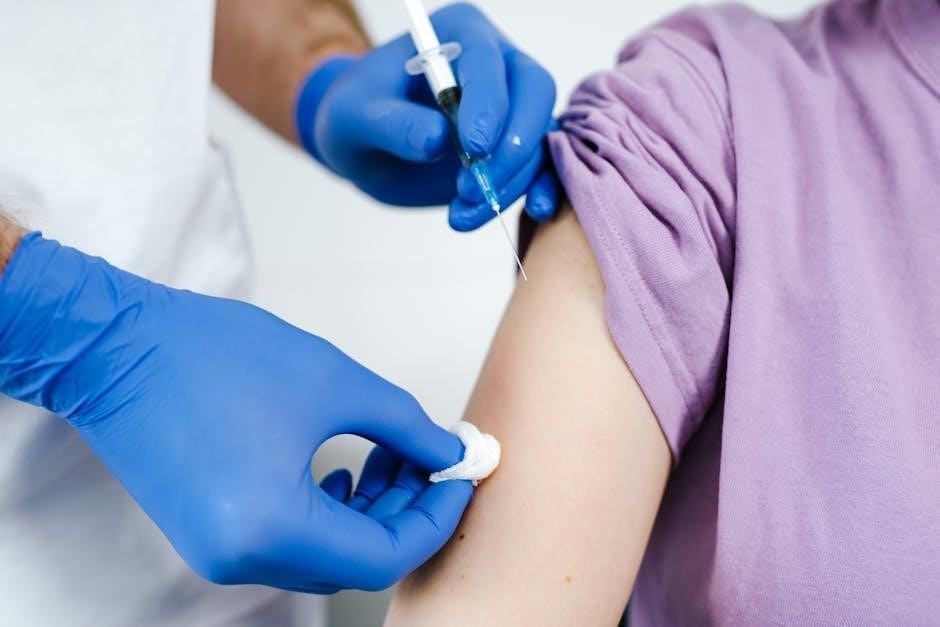
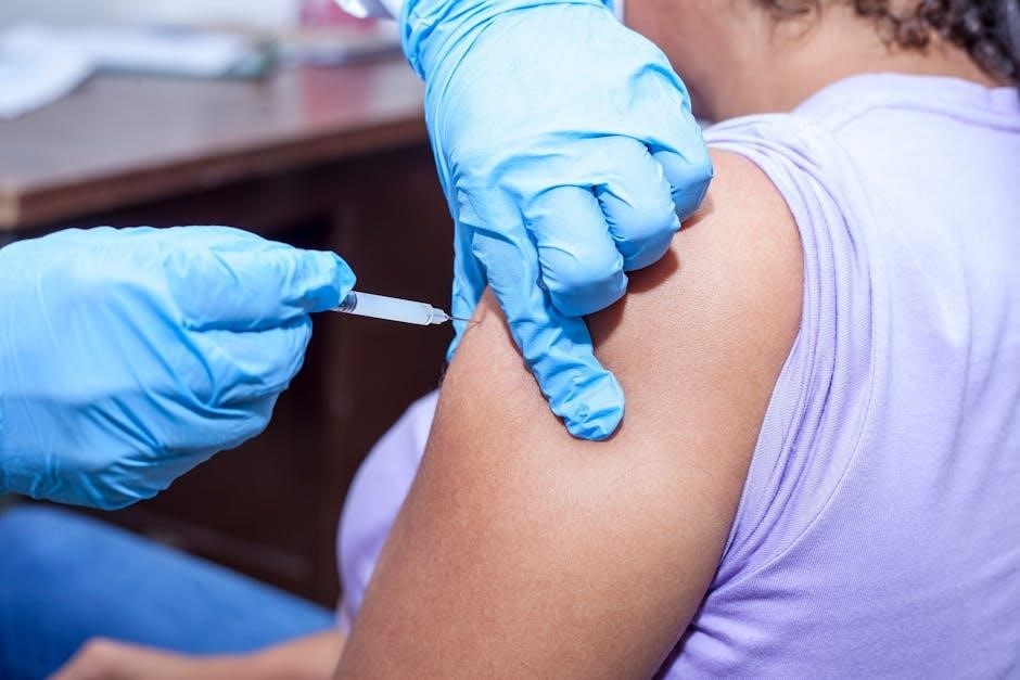
Initially, the patient is positioned to optimize visualization of the target anatomy under the fluoroscope. Sterile preparation of the skin is paramount, followed by a local anesthetic injection to numb the area. A small needle, visible on the fluoroscopic screen, is then carefully advanced towards the intended joint or space.

Throughout the procedure, the physician continuously monitors the needle’s trajectory in real-time, making adjustments as needed. Contrast dye is often injected to confirm accurate placement and spread within the target area. Once optimal positioning is achieved, the therapeutic medication is administered. Post-injection, the needle is removed, and a bandage is applied, completing the guided process.
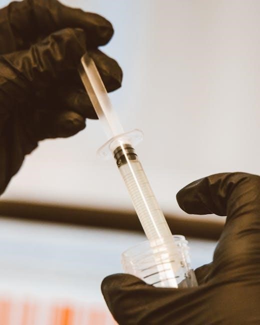
Contrast Dye Usage in Fluoro-Guided Injections

Contrast dye plays a vital role in fluoro-guided injections, enhancing visualization of anatomical structures and confirming accurate needle placement. Injected under fluoroscopic guidance, the dye allows physicians to observe the spread of the medication in real-time, ensuring it reaches the intended target area effectively.
This confirmation is crucial for maximizing therapeutic benefit and minimizing the risk of off-target effects. The dye’s visibility helps identify potential issues like vascular or nerve impingement. While generally safe, potential allergic reactions to contrast dye are considered, and appropriate precautions are taken. Careful monitoring during and after injection is standard practice.
Benefits of Fluoroscopic Guidance
Fluoroscopic guidance significantly improves the precision and safety of injections, leading to enhanced patient outcomes. Real-time visualization allows for accurate needle placement, ensuring the medication reaches the precise anatomical location intended for treatment. This minimizes the risk of injecting into unintended areas, reducing potential complications and maximizing therapeutic efficacy.
Furthermore, fluoroscopy confirms appropriate medication spread, verifying the drug is effectively targeting the source of pain or inflammation. It’s particularly beneficial in complex cases or when anatomical landmarks are difficult to palpate. Ultimately, fluoroscopic guidance contributes to more reliable and consistent injection procedures.
Risks and Potential Complications
While fluoro-guided injections are generally safe, potential risks exist. Radiation exposure, though typically low, is a primary concern, necessitating careful monitoring and minimization techniques. Infection, as with any invasive procedure, remains a possibility, requiring strict sterile protocols. Nerve damage, though rare, can occur if the needle inadvertently contacts a nerve during insertion.
Other potential complications include bleeding at the injection site, allergic reaction to contrast dye or medication, and temporary discomfort. It’s crucial to discuss these risks with your physician before the procedure. Prompt reporting of any unusual symptoms post-injection is essential for timely management of any adverse events.
Radiation Exposure
Fluoroscopy utilizes ionizing radiation to create real-time X-ray images, inherently posing a radiation exposure risk. However, modern fluoroscopic equipment employs techniques to minimize dosage, such as pulsed fluoroscopy and collimation, focusing the beam on the target area. Physicians adhere to the ALARA (As Low As Reasonably Achievable) principle, limiting exposure duration and utilizing lead shielding for both patient and staff.
The cumulative radiation dose from a single fluoro-guided injection is generally considered low, comparable to natural background radiation over a short period. Nevertheless, patients should inform their doctor about any prior radiation treatments or pregnancies to optimize safety protocols.
Infection Risk
While fluoro-guided injections are generally safe, any invasive procedure carries a potential, albeit low, risk of infection. Strict sterile technique is paramount during the procedure, including thorough skin preparation with antiseptic solutions and the use of sterile equipment and gloves. The injection site is carefully monitored post-procedure for signs of infection, such as increasing pain, redness, swelling, or drainage.
Prophylactic antibiotics are typically not routinely administered before fluoro-guided injections, unless the patient has specific risk factors, like a compromised immune system or a history of prior infections. Prompt medical attention is crucial if any infection symptoms develop following the injection.
Nerve Damage
A rare, but significant, complication of fluoro-guided injections is potential nerve damage; This risk arises from the proximity of nerves to the injection site, particularly during spinal or peripheral joint injections. Direct needle trauma or irritation from the injected medication can affect nearby nerve structures, leading to symptoms like pain, numbness, tingling, or weakness in the affected limb.
Fluoroscopic guidance minimizes this risk by allowing precise needle placement, avoiding direct nerve impingement. However, it doesn’t eliminate it entirely. Careful technique, anatomical knowledge, and real-time visualization are crucial. Most nerve injuries are transient, resolving within days or weeks, but permanent damage is possible, though uncommon.
Alternatives to Fluoroscopy
While fluoroscopy offers real-time visualization, alternative image-guidance techniques exist for certain injection procedures. Ultrasound guidance is increasingly popular, particularly for superficial joints like the shoulder or knee, offering a radiation-free option. It’s operator-dependent, requiring expertise in musculoskeletal ultrasound.
CT guidance provides excellent anatomical detail but involves higher radiation exposure than fluoroscopy. It’s often reserved for complex spinal injections or when fluoroscopy is insufficient. Furthermore, fluoroscopy-free Retrograde Intrarenal Surgery (RIRS) is emerging, demonstrating success in select cases, though fluoroscopy should remain readily available for unexpected intraoperative challenges.
Ultrasound Guidance
Ultrasound guidance presents a compelling, radiation-free alternative to fluoroscopy for many injection procedures, especially those targeting superficial structures. Its dynamic imaging capabilities allow real-time visualization of nerves, vessels, and the target joint, enhancing needle placement accuracy. However, ultrasound’s effectiveness is heavily reliant on the operator’s skill and experience in musculoskeletal sonography.
While excellent for visualizing soft tissues, ultrasound’s penetration depth is limited, making it less suitable for deep spinal structures. Despite this, advancements in ultrasound technology and techniques are continually expanding its applications in image-guided injections, offering a safe and effective option for appropriate patients.
CT Guidance
Computed Tomography (CT) guidance offers superior bony visualization compared to fluoroscopy or ultrasound, making it particularly valuable for injections targeting structures closely related to bone, such as facet joints or the sacroiliac joint. CT provides detailed anatomical information in all three planes, allowing for precise needle placement and confirmation of accurate drug delivery.
However, CT involves significantly higher radiation exposure than fluoroscopy, limiting its use to situations where the benefits clearly outweigh the risks. It’s generally reserved for complex cases or when fluoroscopy is insufficient to adequately visualize the target anatomy. Careful consideration of radiation safety protocols is paramount when utilizing CT guidance.
Fluoroscopy-Free RIRS (Retrograde Intrarenal Surgery) Considerations
While traditionally reliant on fluoroscopic guidance, Retrograde Intrarenal Surgery (RIRS) is increasingly performed without it, demonstrating feasibility in select cases. This approach aims to minimize radiation exposure for both patients and medical personnel. However, successful fluoroscopy-free RIRS necessitates experienced surgeons with a thorough understanding of renal anatomy and meticulous surgical technique.
It’s crucial to acknowledge that unexpected intraoperative challenges can arise, potentially requiring a conversion to fluoroscopic guidance. Therefore, maintaining fluoroscopy availability during the procedure is highly recommended, ensuring adaptability and optimal management of unforeseen circumstances, ultimately prioritizing patient safety and surgical success.
Patient Preparation for a Fluoro-Guided Injection
Prior to a fluoro-guided injection, a comprehensive medical history review is essential, including allergies, medications (especially blood thinners), and prior imaging results. Patients should inform their physician of any possibility of pregnancy. A detailed discussion regarding the procedure, its benefits, and potential risks is paramount, ensuring informed consent.
On the day of the procedure, patients may be asked to abstain from food and drink for a specified period. They should wear loose, comfortable clothing. Depending on the injection site, specific preparation may be required, such as shaving the area. Vital signs will be monitored, and an IV line may be inserted for medication administration, optimizing patient comfort and safety throughout the process.
Post-Injection Care and Recovery
Following a fluoro-guided injection, patients are typically monitored for a short period to observe for any immediate adverse reactions. Pain levels should be regularly assessed, and any significant increase reported promptly. Activity restrictions are common, varying based on the injection site and individual needs; avoid strenuous activity initially.
Patients are advised to follow their physician’s specific instructions regarding wound care, medication usage, and follow-up appointments. Ice packs can help manage discomfort and swelling. While some temporary soreness is expected, persistent or worsening pain warrants medical attention. Hydration is also encouraged to aid in recovery and overall well-being.
The Future of Image-Guided Injections
The evolution of image-guided injections is leaning towards minimizing radiation exposure and enhancing precision. Advancements in artificial intelligence (AI) and machine learning are poised to refine image analysis, improving needle guidance and target accuracy. Research continues to explore alternative imaging modalities, like advanced ultrasound techniques, to reduce reliance on fluoroscopy.

Furthermore, the development of novel contrast agents with improved visibility and biocompatibility is anticipated. Robotic-assisted injection systems could offer enhanced stability and control. Ultimately, the goal is to deliver safer, more effective, and personalized pain management solutions through increasingly sophisticated image-guided techniques.